Herpes zoster ophthalmicus — shingles affecting the eye. How the herpesvirus family (VZV vs HSV, chickenpox vs shingles) differs, where it strikes (eyelid skin, cornea, or orbit), urgent antiviral treatment, and oculoplastic repair of the eyelid scarring it leaves behind.
Medically reviewed by EyePlastics Medical Editorial BoardASOPRS oculoplastic surgeonsLast updated June 2026
“Shingles in the eye” — medically herpes zoster ophthalmicus (HZO) — happens when the chickenpox virus, dormant for decades in a nerve near your brain, reactivates along the nerve that supplies the forehead, upper eyelid, and eye. It causes a painful, blistering rash on one side of the face and can threaten the eye itself. Because several different viruses in the “herpes” family affect the eye in different ways — some the skin, some the cornea, some even the orbit — the names are genuinely confusing. This guide untangles them and explains what shingles around the eye means for your vision.
This is an in-depth companion to our main Eyelid & Orbital Infections guide. Shingles affecting the eye is an urgent problem — antiviral treatment works best when started within 72 hours of the rash appearing (and may still help beyond that window if new blisters are still forming or the eye is involved).
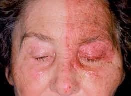
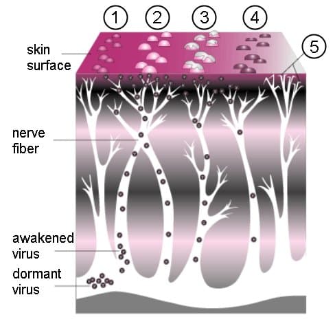
Two members of the human herpesvirus family cause most eye disease, and each has more than one name depending on how and when it strikes. This is the single biggest source of confusion, so here it is laid out plainly:
| Virus | Names you may hear | How it behaves around the eye |
|---|---|---|
| Varicella-zoster virus (VZV) | Chickenpox (first infection) · Shingles / herpes zoster (reactivation) · HZO when it hits the eye nerve | One virus, two illnesses at different times of life. Childhood chickenpox, then decades later the same virus can reawaken as shingles in a single nerve territory. |
| Herpes simplex virus (HSV) | HSV-1 (cold sores, most eye cases) · HSV-2 (usually genital) · Herpetic eye disease / herpes keratitis | A different virus that tends to recur on the same eye. Its signature problem is a branching (“dendritic”) ulcer on the cornea — not the dermatomal facial rash of shingles. |
The key distinction: varicella is the first-ever VZV infection (chickenpox, usually in childhood, a widespread rash). Herpes zoster (shingles) is the reactivation of that same virus years later, confined to one nerve’s strip of skin. When that nerve is the ophthalmic division of the trigeminal nerve, you get herpes zoster ophthalmicus. HSV is an entirely separate virus that most often troubles the cornea and comes back repeatedly.
Patients are often told they have “herpes in the eye” and understandably worry — but the location matters enormously, because each site is a different problem managed by a different part of eye care. Viral eye disease shows up in three broad zones:
1. Eyelid & periocular skin
The dermatomal blistering rash of shingles, and later its scarring — cicatricial ectropion, eyelid retraction, ptosis, notching, and lash loss. This is the oculoplastic surgeon’s territory: reconstructing the eyelid so it protects the eye again.
2. Cornea & ocular surface
Both viruses can inflame the front of the eye — HSV classically as a branching dendritic ulcer, VZV as pseudodendrites, stromal keratitis, uveitis, and a numb (“neurotrophic”) cornea. This is sight-threatening and managed closely by ophthalmology/cornea specialists.
3. The orbit (eye socket)
Uncommon but serious: VZV can inflame the deep structures behind the eye — the muscles and nerves — producing an orbital-inflammatory picture with double vision, a droopy lid, or optic-nerve involvement (orbital apex syndrome). See Orbital Conditions.
So “shingles of the eye” can mean a rash on the lid, an ulcer on the cornea, or inflammation deep in the socket — often more than one at once. That is why anyone with shingles in the eye region should be examined by an eye specialist, not treated on the rash alone.
Shingles around the eye usually announces itself before the rash: a few days of burning, tingling, or aching pain on one side of the forehead, sometimes with fever or malaise. Then the hallmark rash appears:
HZO can cause trouble both during the acute illness and long after the rash heals:
Oral antiviral therapy — acyclovir, valacyclovir, or famciclovir — should ideally be started within 72 hours of the rash appearing to shorten the illness, lessen its severity, and reduce the risk of postherpetic neuralgia. When the eye is involved, an ophthalmologist co-manages the cornea and any internal inflammation with topical and sometimes additional systemic therapy. Later, once the infection has fully settled, an oculoplastic surgeon can repair the cicatricial eyelid problems shingles leaves behind — correcting ectropion, retraction, or ptosis so the eyelid protects the eye again. Any red, painful, or light-sensitive eye during shingles warrants same-day evaluation.
Prevention: The Shingrix vaccine (two doses) is recommended for adults aged 50 and older regardless of prior shingles history, and for immunocompromised adults aged 19 and older who are at higher risk. It lowers the overall risk of shingles by more than 90% (about 97% in adults 50-69 and about 91% in those 70 and older) and substantially reduces the risk of postherpetic neuralgia — the most effective way to avoid HZO in the first place.
Treat shingles anywhere near the eye as urgent. Seek same-day care if you develop a one-sided facial rash with any eye redness, pain, light sensitivity, or change in vision — and especially if blisters appear on the nose (Hutchinson’s sign). Early antivirals plus an eye examination protect vision in the acute phase; an oculoplastic surgeon addresses the eyelid scarring and malposition that can follow.
Shingles near the eye needs specialist eyes on it
From acute antiviral care to repairing eyelid scarring afterward, HZO is best managed by an eye specialist. Find an ASOPRS-trained oculoplastic surgeon near you.
Connect with a board-certified oculoplastic surgeon who specializes in shingles in the eye (herpes zoster ophthalmicus).
Search the Directory →Oculoplastic Surgery — Patient Education
Anophthalmos